

Eye Socket
Layer 1
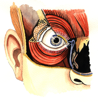
Under the skin around the eye there is a circle of muscles which constrict, to close the eye lids and bring the eye brow down, to protect the eye ball.
Near the top and outer side of the eye the tear producing gland, (Lacrimal gland), makes tears to wash across the eye. These are drained away down fine canals, (in blue), to drain into the airway of the nose. This is why our nose runs when we cry.
Layer 2
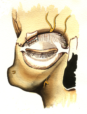
Under the muscles there is a partition which walls off the eye socket, it is continuous with the lids.
Nerves from deeper in the socket go to the muscles and skin.
Layer 3
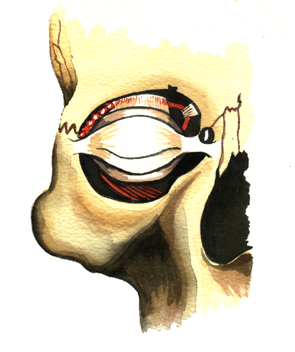
The eye lids are attached to either side of the bony socket. The eye ball is behind the lids.
With the socket partition removed, some of the eye’s muscles can be seen.
Layer 4
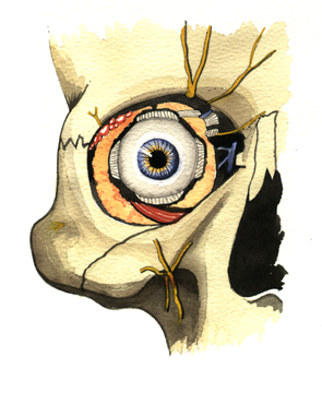
Behind the socket partition and lids, the eye ball floats in a cushion of fluid fat. It is moved by the eye muscles.
Eyeball Exterior
Layer 5
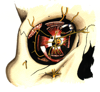
If the eyeball is removed muscle stumps and nerves and tear gland (and lids) remain.
Layer 6
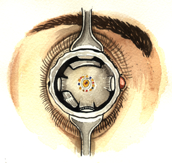
This would be the surgical view, before the transparent socket skin is closed to seal it.
Layer 7
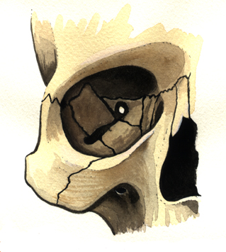
There are many bones that make up the eye socket. They are easily broken by a punch or kick or squash ball.
Sometimes bones are removed to create space around a swollen eye, (occasionally to treat thyroid eye disease).
This information site has been provided by varied UK and US eye doctors for patients with eye problems.
Once your eye doctor has made a diagnosis or recommended an investigation or treatment, then you will be able to find further explanation on this site.
It is not a self diagnosis centre. It should not be relied upon without taking professional advice.