

Eyeball Workings
Aqueous Flow
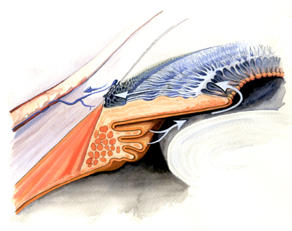
The continuous formation of internal ‘Aqueous’ fluid, by the ciliary body epithelium, keeps the eye ball inflated.
The fluid circulates from behind the iris, over the lens through the pupil around the front chamber of the eye, over the cornea and filters out through a meshwork to drain into veins and return to the heart.
Accommodation
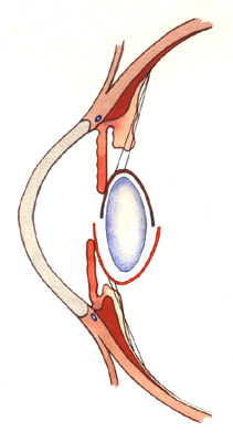
This occurs when the eye changes focus from looking at an object in the distance to reading a book, (or looking at another near object).
The ciliary body is the eye muscle responsible for changing the shape and position of the eye lens.
The Zonules are very fine fibres that suspend the lens from the ciliary body ring, just behind the iris and pupil.
The diagram is in 2 parts;
Top: the circular muscle (dark brown) is relaxed, (back against the eye wall), this puts tension on the fibres which stretch the lens into a flatter disc. (Less powerful focus)
Bottom: The Circular ciliary muscle contracts towards the centre of the eye. This slackens the zonule fibres, then the lens springs back into a rounder shape, which has greater focussing power.
Presbyopia
This is a normal loss of the ability to focus on near objects, such as reading - see Accommodation.
This usually occurs in mid life, (in the 40’s), initially it shows itself when someone holds the news paper further away to focus on it. Eventually this trick doesn’t compensate and reading glasses are required. Short sighted people often experience near reading difficulties a few years later.
This information site has been provided by varied UK and US eye doctors for patients with eye problems.
Once your eye doctor has made a diagnosis or recommended an investigation or treatment, then you will be able to find further explanation on this site.
It is not a self diagnosis centre. It should not be relied upon without taking professional advice.